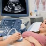
Pregnancy is a time for anticipation, hopes, and myriad emotions. For most expectant parents, the entire journey goes on smoothly and without any hiccups. Sometimes, however, pregnancies become “high-risk,” necessitating closer monitoring to ensure the welfare and survival of both mother and baby. In these situations, pregnancy ultrasound scans may bring in valuable information-later and life-saving-extraordinaire.
Since then, ultrasound technology has gained much momentum. But its revolution is from those simple and flat two-dimensional images to now intricate and powerful 3D and even 4D scans, allowing parents a live view of the growing baby within the womb itself. You can hardly call this just a pictorial view of the womb in cases of high-risk pregnancy. Rather, it is a real-time investigation of the child’s current status along with treatment and monitoring, allowing the physician to make well-informed decisions at every instance.
What Constitutes a High-Risk Pregnancy?
High-risk pregnancy occurs when there is an increased likelihood of complications on the mother‘s, baby‘s, or both‘s health. High-risk pregnancies can include:
Advanced maternal age (older than 35)
Pre-existing medical conditions like diabetes, high blood pressure, or thyroid disease
Multiple gestation (twins or more)
Pregnancy complications or miscarriage history
Placental issues
Foetal abnormalities or growth limitations
Infection or exposure to toxins during pregnancy
Monitoring both early and continuously is important in high-risk pregnancies to monitor foetal development, identify complications at an early stage, and prepare for required interventions.
The Fundamental Role of Ultrasound in High-Risk Pregnancies
Ultrasound is a non-invasive, safe, and widely used imaging method in prenatal care. It works by using sound waves to create real-time images of the baby, placenta, and uterus. In high-risk pregnancies, ultrasounds are used more frequently and with a more specific diagnostic purpose. Here’s how they contribute at various stages:
1. Confirming Viability and Dating the Pregnancy
The first trimester scan, often performed between 6 to 10 weeks, helps confirm whether the pregnancy is viable (presence of heartbeat) and whether it is located correctly within the uterus. In high-risk pregnancies, early confirmation is crucial, especially if the mother has a history of ectopic pregnancies or miscarriages.
This scan also helps accurately date the pregnancy, which is essential for tracking growth later on.
2. Nuchal Translucency and First Trimester Screening
Between 11 and 14 weeks, the nuchal translucency scan is done to assess the risk of chromosomal abnormalities like Down syndrome. This is especially relevant for older mothers or those with a family history of genetic conditions.
Ultrasound in this stage provides measurements that, when combined with blood tests, help determine whether further testing such as CVS or amniocentesis is needed.
3. Detecting Structural Anomalies in the Second Trimester
The detailed anomaly scan, typically done at 18–22 weeks, is one of the most important scans in a high-risk pregnancy. It provides a comprehensive look at the baby’s anatomy — including the brain, heart, spine, kidneys, and limbs.
Any potential structural abnormalities can be detected, and a care plan can be put in place early. In cases where foetal surgery may be required after birth, this scan allows the medical team to coordinate appropriately.
In some instances, parents may opt for an Early Gender Scan, often around 16 weeks, to learn the baby’s sex. While gender identification is a personal choice, in high-risk cases involving sex-linked genetic disorders, this information can become clinically relevant.
4. Monitoring Growth and Amniotic Fluid Levels
One of the key concerns in high-risk pregnancies is intrauterine growth restriction (IUGR), where the baby isn’t growing as expected. Serial growth scans are used to track foetal weight, abdominal circumference, and head size.
Ultrasound is also used to measure amniotic fluid levels. Too little (oligohydramnios) or too much (polyhydramnios) fluid can indicate problems such as placental insufficiency, gestational diabetes, or foetal distress.
5. Assessing Placental Position and Function
The placenta plays a vital role in nourishing the baby. In high-risk pregnancies, issues like placenta previa (where the placenta covers the cervix) or placental abruption (where it detaches prematurely) can be dangerous. Ultrasound scans help monitor the position and function of the placenta throughout pregnancy.
Doppler ultrasound, a specialised form, is often used to evaluate blood flow in the umbilical cord, placenta, and baby’s brain. This is particularly important in managing conditions such as preeclampsia or when there is reduced foetal movement.
6. Planning for Delivery
As the due date approaches, ultrasound helps determine the baby’s position (breech, transverse, cephalic), weight estimate, and readiness for birth. In high-risk cases, this information is essential for planning whether a vaginal delivery is safe or a caesarean section is required.
For mothers with previous caesarean sections or uterine surgery, ultrasound can check the thickness of the uterine scar to assess the risk of rupture during labour.
7. Guiding Medical Procedures
Invasive procedures such as amniocentesis or chorionic villus sampling (CVS) are sometimes needed in high-risk pregnancies. These are performed under ultrasound guidance to ensure precision and reduce risk.
Ultrasound also plays a role in procedures like intrauterine transfusions, foetal shunt placements, or selective reduction in multiple pregnancies.
Psychological Reassurance and Parental Bonding
Beyond the medical benefits, regular ultrasound scans in pregnancy offer immense emotional reassurance for expectant parents. For those navigating the uncertainty of a high-risk pregnancy, each scan offers a sense of connection and progress.
Even in medically complex cases, being able to see their baby moving, growing, and developing can strengthen parental bonding. Many clinics now offer 3D and 4D scans, which provide even more detailed visualisation and help parents feel closer to their unborn child.
Customized Ultrasound Schedules in High-Risk Cases
In contrast to low-risk pregnancies where only a few routine scans are needed, high-risk pregnancies can need a tailored ultrasound regimen. This is based on the underlying condition and upon assessment by the obstetrician. A few women may undergo fortnightly or even weekly scans during the third trimester in order to monitor their baby’s health closely.
Coordination among sonographers, obstetricians, and maternal-foetal medicine specialists also means that no detail goes unnoticed.
Technology That Saves Lives
Modern ultrasound technology is increasingly becoming more precise, portable, and accessible. Innovations like high-resolution transducers, Doppler capabilities, and AI-assisted image analysis are enhancing the accuracy of diagnoses.
Moreover, telemedicine and cloud-based imaging platforms are allowing faster sharing of results between specialists, ensuring timely interventions in critical situations.
When to Be Proactive
If you’re pregnant and have been told your pregnancy is high-risk — or you suspect it might be — don’t hesitate to ask your healthcare provider about additional ultrasound monitoring. Early intervention is one of the best ways to reduce the chances of complications and improve outcomes.
Similarly, if you have specific questions about a baby’s gender, growth, or development, services like an Early Gender Scan can complement your medical care by offering clarity at earlier stages.